In radiology, we are concerned with the application of imaging techniques for the diagnosis and treatment of diseases. These include X-ray, CT, MRI and ultrasound, among others. Radiology enables rapid and accurate diagnosis of various diseases and injuries. The procedures are gentle and in many cases an alternative to invasive diagnostics.
Our partner institute in the Medical Health Center is the Radiologie Südost Institute. The institute is made up of a group of medical specialists organised in the form of a partnership for medical imaging. It offers a comprehensive range of state-of-the-art imaging services with MRI including neuro-MRI, X-ray, ultrasound, computed tomography, mammography and osteodensitometry.
What we offer
Computed tomography (CT) is a sectional imaging technique and a further development of X-ray technology. It is mainly used to examine the head, lungs, abdominal organs, spine and bones.
How does CT work?
The procedure involves circling an X-ray tube around the body on a ring and generating a narrow X-ray beam. This beam penetrates the desired part of the body, and is attenuated to varying degrees by the various structures (e.g. fat, muscles, organs, bones). Exactly opposite the X-ray tube on the same ring is a wide range of sensors (detectors) that receive the attenuated signal. CT provides a very detailed, multidimensional view of the inside of the human body.
Bone density measurement (bone densitometry) measures the density and the calcium salt content of bones. It is used to detect osteoporosis and monitor its treatment.
How does a bone density measurement work?
For the examination, X-rays of very low intensity are sent through the body or through the bones, and the attenuation of the rays is measured, making it possible to determine the bone mass. The result can be used to infer the mineral content of the bone and thus its density. Reduced bone density indicates a risk of increased bone fragility. The lumbar spine and the hips are used as measurement locations, with a level of radiation exposure that is so low that it is negligible.
Magnetic resonance is considered the best method available for examining the brain, the spinal column, the joints and soft tissue. MRI (magnetic resonance imaging) is a modern tomographic imaging technique that can generate images of any area of the body.
How does a magnetic resonance imaging work?
It does not use X-rays for this, but rather a strong magnetic field and radio waves. The signals received are processed by a computer, with the resulting images allowing for an extremely accurate distinction between normal and abnormal changes in the body, especially in soft tissue such as the brain or internal organs.
Mammography is a special X-ray examination of the breast and is used in breast cancer screening – the earlier the disease is recognised, the better are the chances of recovery.
How does a mammography works?
Mammography isis carried out on special X-ray machines using “soft” X-rays that result in higher-contrast images. Fine differences in the density and composition of the tissue can be seen in the images. Even microscopic calcifications (microcalcifications), which are often a first indication of breast cancer, can be detected.
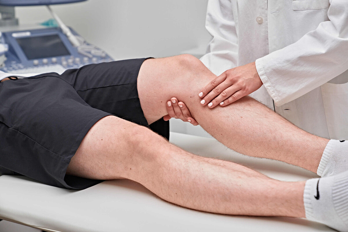
Ultrasound technology is used above all to examine abdominal and pelvic organs (such as the liver, gall bladder, pancreas, spleen, kidneys and bladder) and soft tissues (including the thyroid gland, breast), joints and vessels.
How does ultrasound scans work?
The examination is carried out without the use of X-rays. High-frequency sound waves (ultrasound) that are inaudible to the human ear are reflected in different ways by the various organs. The transducer receives the reflected sound waves, converts them into electrical impulses and transmits them to the ultrasound machine, where the impulses are amplified and displayed on a screen. Using special colour Doppler (duplex) sonography procedures, the examiner can obtain additional information about the speed and direction of blood flow in vessels. This can be used to detect vascular diseases such as vasoconstriction in arteriosclerosis or vein occlusion (thrombosis).
Conventional X-ray imaging is generally used for an initial assessment of bones, the lungs and the heart. Exposing an X-ray film takes only fractions of a second.
Our team of experts


Contact and consulation
Make an appointment and we will be happy to advise you on our offer.
+41 81 303 38 61
radiologie.ragaz@hin.ch